What's New
(20230529) We are finally back in the busines of releasing data. To subscribe to our resources, please go here and follow the instructions carefully.
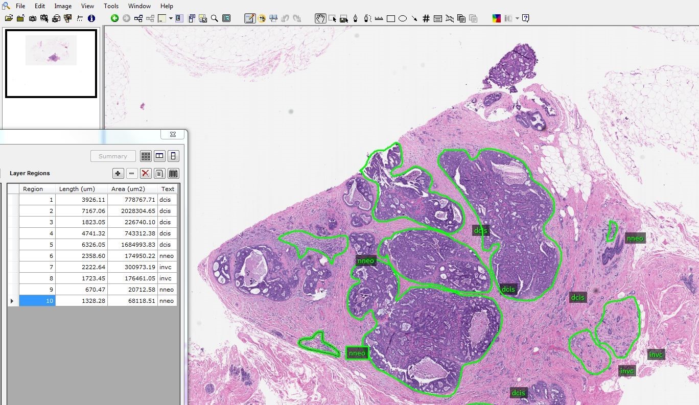
(20220113) We are pleased to announce v2.0.0 of the TUH DPATH Corpus. This release contains a breast tissue subset and is described here. There are 3,505 annotated images partitioned into training, development and blind evaluation sets.
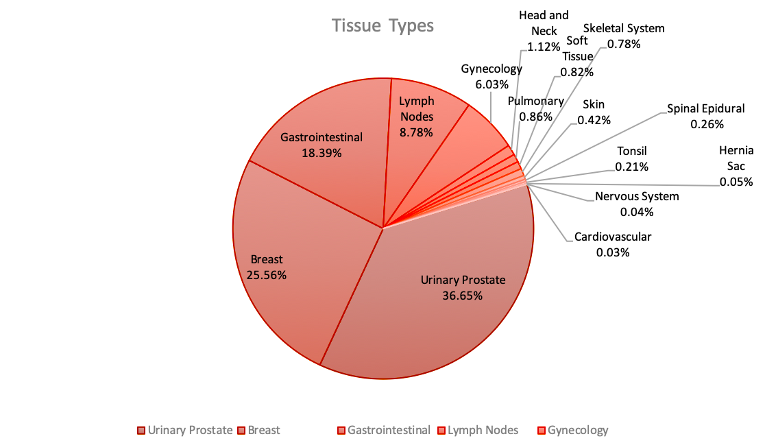
(20211215) We have completed scanning the Fox Chase Cancer Center subset and now have scanned over 80,000 slides.
Project Summary
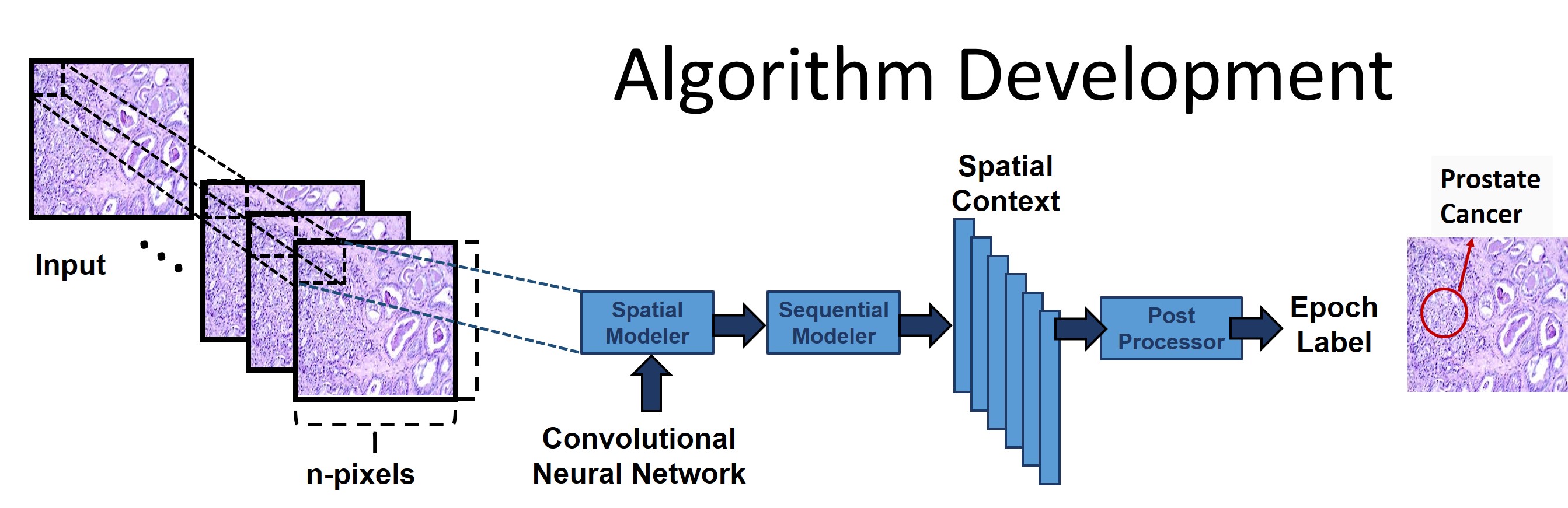 In this NSF-funded project, we are developing a digital
imaging system using big data and machine learning algorithms
to automatically characterize pathology slides. We have
developed a sustainable facility to rapidly collect
automatically annotated whole slide images. This project is
producing the necessary data resources to support the
development of high performance deep learning models.
In this NSF-funded project, we are developing a digital
imaging system using big data and machine learning algorithms
to automatically characterize pathology slides. We have
developed a sustainable facility to rapidly collect
automatically annotated whole slide images. This project is
producing the necessary data resources to support the
development of high performance deep learning models.
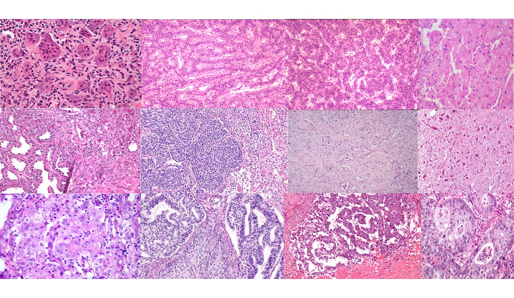
Over 10M slides are read each year in the U.S. alone. Tapping
into a fraction of this data will allow significant
advancement of the science. Healthcare providers and machine
learning researchers will be able to access an open source
high-quality searchable archive of clincial data. More
information on this project can be found
here.
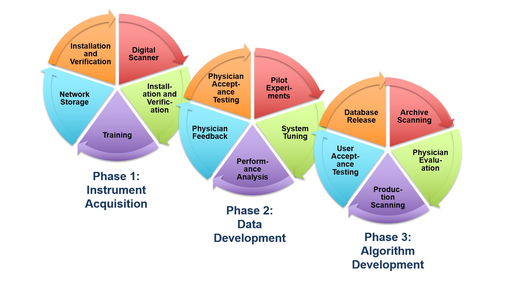
A Cost-Effective Image Management Platform
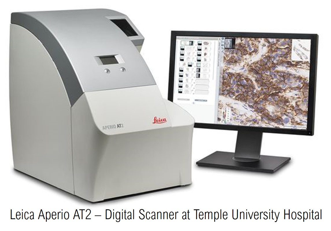
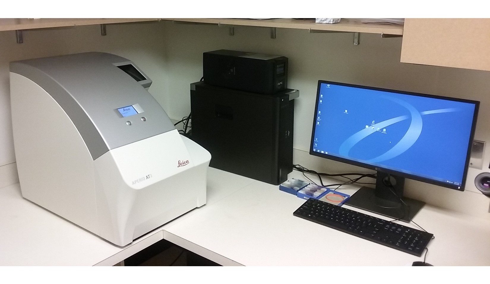
This NSF Major Research Instrumentation (MRI) grant supported
the purchase of a
Leica Aperio AT2
scanner as the platform used to convert pathology slides to
digital images. This scanner can scan 50 high quality TIFF images
with lossless compression per hour.
We have also developed a very cost-effective Petabyte file store
based on off-the-shelf components to store our database of 1M
pathology images. To learn more about our clustered computing
environment being developed to support this research program,
read this
overview.